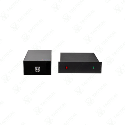
Quantitative Kerr Microscopy
Quantitative Kerr microscopy measures the magnetization direction and magnitude at specific points or across the surface of magnetic materials using the magneto-optical Kerr effect — converting polarization rotation data from reflected light into calibrated magnetization maps rather than qualitative domain images. This quantitative capability gives materials scientists and magnetic device researchers direct access to the physical magnetization vector at each measurement point rather than relative contrast information alone.
The measurement principle reflects polarized light from the sample surface. Magnetized regions rotate the polarization plane of reflected light by a Kerr angle proportional to the local surface magnetization component. A calibrated polarimeter setup measures this rotation angle precisely at each point — using a photoelastic modulator or Faraday cell to modulate the polarization state and lock-in detection to extract the rotation signal from background optical noise. Furthermore, three geometric configurations target different magnetization components. Polar Kerr geometry detects out-of-plane magnetization. Longitudinal geometry detects in-plane components parallel to the plane of incidence. Transverse geometry detects in-plane components perpendicular to the incidence plane.
Quantitative conversion from measured Kerr rotation angle to magnetization magnitude requires calibration against a material with known saturation magnetization. Calibration establishes the instrument’s sensitivity in degrees per unit magnetization — allowing subsequent measurements to report local magnetization in absolute units of A/m or Tesla rather than arbitrary contrast units. Additionally, scanning Kerr microscopy systems raster-scan the focused laser spot across the sample surface with motorized XY stages. They build complete magnetization maps with spatial resolutions approaching the optical diffraction limit at 0.5 to 2 µm.
Time-resolved Kerr microscopy adds stroboscopic pump-probe capability. Ultrashort laser pulses pump a magnetic switching event. Delayed probe pulses capture the magnetization state at precise time intervals after excitation. As a result, magnetization dynamics on picosecond to nanosecond timescales become directly measurable — covering spin wave propagation, domain wall motion, and ultrafast demagnetization processes relevant to next-generation magnetic memory and spintronic device development.
These systems serve university physics and materials science departments in Lahore and Karachi studying spintronic device physics, magnetic recording media development programs, defense materials research organizations, and pharmaceutical nanotechnology research groups investigating magnetically functionalized particle systems for drug delivery applications.
Showing the single result