Stereoscopic Ophthalmic Microendoscope System
A Stereoscopic Ophthalmic Microendoscope System is a highly specialized surgical visualization tool used in delicate eye surgery. It combines a very small diameter rigid endoscope (often <1mm) with a stereoscopic (3D) camera system. This allows ophthalmic surgeons to view and navigate within the intricate structures of the eye—such as the retina or anterior chamber—through extremely small incisions, providing magnified, high-resolution, depth-perceptive imagery that is critical for procedures like vitrectomy or micro-stent placement. This technology enhances surgical precision, reduces tissue trauma, and improves outcomes in minimally invasive ophthalmic surgeries.
-
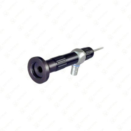
Ultra-thin rigid endoscope (<1mm) for access through minute incisions in the eye.
-
Provides true stereoscopic (3D) high-definition video for depth perception during microsurgery.
-
Used in retinal surgery, glaucoma surgery, and other complex intraocular procedures.
-
Enables surgeons to see around corners and into spaces not visible with operating microscopes alone.
-
Integrates with surgical illumination and recording systems.
-
Represents the cutting edge of minimally invasive ophthalmic surgical technology.
See in 3D inside the eye. The Stereoscopic Ophthalmic Microendoscope System enables unparalleled precision in minimally invasive eye surgery.
Showing the single result